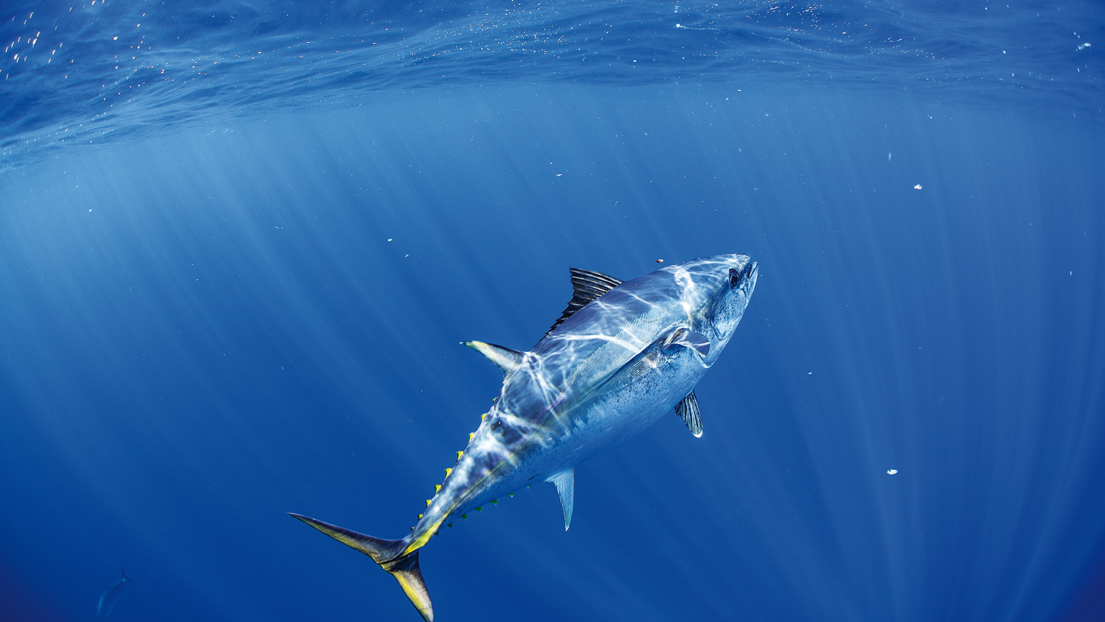

The use of fish cell lines, both as a research tool and a diagnostic tool, has played a major role in the development of salmonid and cyprinid aquaculture worldwide. The commercial success of these finfish aquaculture industries is due, in part, to the development of fish cell lines which are used to monitor farmed fish populations for the presence of specific viral pathogens. Based on the results of such health surveillance programs disease-free stocks can be kept isolated from infected stock through restrictions in fish movements. The current lack of continuous tuna cell lines suitable for the isolation and growth of viral pathogens of tuna could be a serious obstacle to effective disease control in tuna hatcheries and nurseries which, in turn, could have a significant negative impact on the future development of the tuna aquaculture sector. It is noteworthy that viral infections of a tuna species (Thunnus thynnus) have been documented (1). Moreover, other viral pathogens such as marine nodaviruses (2) and birnaviruses (3) tend to be catholic in their host range and should be considered a significant risk.
Development of diagnostic tools for identification of viral pathogens in other systems has been reliant on the availability of continuous cell lines for virus cultivation. Isolation and growth of viral pathogens in susceptible cell lines provide an almost limitless supply of partially purified virus for the development of improved diagnostic procedures for these pathogens (4). In order to be able to develop similar systems to service the farmed tuna sector, there is a need for continuous tuna cell lines.
The aim of this project is to develop continuous tuna cell lines to improve our capacity to isolate and characterise tuna viruses, and to enhance our response to new pathogens that may threaten farm production. Identification of disease-free broodstock, eggs and fry is essential for the further development of the tuna aquaculture sector.
REFERENCES
1. Matsuoka S, Inouye K & Nakajima K. 1996. Cultured fish species affected by red sea bream iridoviral disease from 1991 to 1995. Fish Pathol. 31: 233-234.
2. Nishizawa T, Furuhashi M, Nagai T, Nakai T & Muroga K. 1997. Genomic classification of fish nodaviruses by molecular phylogenetic analysis of the coat protein gene. Appl. Environ. Microbiol. 63: 1633-1636.
3. Reno PW. 1999. Infectious pancreatic necrosis and associated aquatic birnaviruses. In: Fish Diseases and Disorders vol. 3 Viral, Bacterial and Fungal Infections (Woo PTK & Bruno DW, eds.) CABI Publishing, New York, NY, Pp. 1-55.
4. Crane MStJ & Bernoth, E-M. 1996. Molecular Biology and Fish Disease Diagnosis: Current Status and Future Trends. Recent Advances in Microbiology 4: 41-82.